Quiesce: Some cells rest. Some stop for good.
qui·esce /kwee-ESS/ verb, of a cell: to rest from dividing, and still be able to start again.
The name is set from seven cells in Figure 1. The paper below is set on the same clock.Set from seven cells in Fig. 1
Quiesce is a research laboratory in Adelaide. We study how cells age, and we are asking why one cell rests and wakes again while its neighbour stops dividing for good. This page is set like a paper, and on a clock: its headings grow wider and thinner as you read down, the way the cells in Figure 1 do when they stop. Its Results section is still blank.
Keywords cell division · quiescence · senescence · tissue · protein shape
Read the abstract ↓ Write to the lab

Drag across the figure to stain cells.
Stain mode is on: the page won’t scroll while your finger is on the figure.
A still of the figure, midway on its clock: the sheet is full, many cells are resting for lack of room, and the first large, flat stopped cells sit in the middle.
§0 · F1 of 10 · Early
What is this lab, in plain words?
Most cells in a body are not dividing at any given moment. Many are resting, and can start again when there is a wound to close or tissue to replace. Some stop for good: they grow large and flat, stay where they are, and change the signals they send to the cells around them. We study that second kind of stopping, in cells grown in dishes and in simple computer models like Figure 1: what tips a cell into it, why neighbours in the same dish behave so differently, and what a stopped cell does to the tissue around it. The questions we can’t answer yet are in §5.
What this lab is not No products. No treatments. No trials. No health claims. Research only.
List of figures
- Fig. 1A sheet of cellsA still
- Fig. 2An alpha helixA still
- Fig. 3ResultsNot drawn yet
- Fig. 4The same sheet, laterA still
- Plates A–EThe lab at workPhotographs
§1 · F2 of 10 · Early
What is the difference between resting and stopping?
Most of the cells in a body are not dividing at any given moment. Many are quiescent1: resting, with the means to divide switched off but kept, ready to start again when there is a wound to close or tissue to replace. We named the lab for that state, because it is the one we most want to understand.
In plain words
1 Quiescent: resting. Not dividing, but able to.
Some cells leave it and don’t come back. They become senescent2: they stop dividing for good, grow large and flat, and change the signals they send to the cells around them. That may be part of how a body keeps a damaged cell from copying itself. But stopped cells gather with age, and nobody yet knows exactly why a cell takes that turn, or whether it is one decision or many small ones.
2 Senescent: stopped. Not dividing, and not going to.
Every cell that divides goes round the cell cycle3. We have set this page to show the two ways a cell can leave it. Below, the word cell is drawn at six moments on Figure 1’s clock: narrow and firm while a cell can still divide, wide and thin once it has stopped. Every heading on the page is set at one of those moments, one step later for each section. The step with the question mark is where a cell turns, and it is what we are trying to draw properly.
3 Cell cycle: the steps a cell goes through to copy itself and split in two.
The word cell, set six times: narrow and firm at frame 0, wider and thinner at frames 2, 4, 6 and 8, and wide and hairline at frame 10.
The shapes · one cell, twice
The shading
- Dividing
- Resting, no room
- Stopping
- Stopped
The shading is ours, drawn for this page. Real signs that a cell has stopped are several, and none of them is a colour. The model also leaves out a great deal, including cells dying.
The stain
Dragging across Figure 1 marks the cells you touch, the way a dye marks cells in a dish. A marked cell passes the mark to both of its daughters when it divides, so a line of cells can be followed through the clock. That is why a real stain is used: to follow a cell and its daughters, and see which of them stop.
The letter register · Fig. 1a, now
- QResting
- UResting
- IResting
- EStopped
- SStopped
- CResting
- EResting
A table of the figure, not of data: each letter of our name is tied to one cell in Figure 1, and set as wide as that cell has stopped.
§2 · F3 of 10 · Early
What does a protein look like up close?
Proteins do much of a cell’s work, and their shape is what lets them do it. Each is a chain of amino acids4 folded into a shape, and one of the commonest shapes is the alpha helix5, a chain wound into a spring and held there by hydrogen bonds6.
In plain words
4 Amino acid: one building block of a protein.
5 Alpha helix: a chain wound into a spring.
6 Hydrogen bond: a weak link that holds one turn to the next.
An ageing cell may keep fewer of its proteins in good shape. Which ones, and whether it matters, is one of the things we ask about.

The chain itself: the same three atoms repeated, one building block after another. Everything else hangs off it.
One amino acid. A protein is a string of these; this helix is 18 of them.
The part of each building block that differs from one kind to the next. Here they are short stubs, because this is a generic helix.
Weak links between each building block and the one four along. They hold each turn to the next and keep the spiral a spiral.
Nothing you could touch. It is how people draw the path of the backbone so the shape reads at a glance.
Seen from a three-quarter view, at rest, with the far side gently out of focus.
Drag, or use the arrow keys, to turn it. Click it, then scroll or pinch to zoom.
§3 · F4 of 10 · Midway
How do we look at a cell?

-
3.1 Growing cells
We grow cells in shallow dishes, in a warm, humid cabinet kept at body temperature. They are fed, moved to new dishes when they run out of room, and checked every morning.
What it can’t tell us A dish is not a body; cells on plastic behave differently from cells in tissue.
-
3.2 Watching them
We photograph the same dishes through a microscope over days and weeks, so we can follow one cell from picture to picture, like a slow film.
What it can’t tell us A picture shows shape, not reasons.
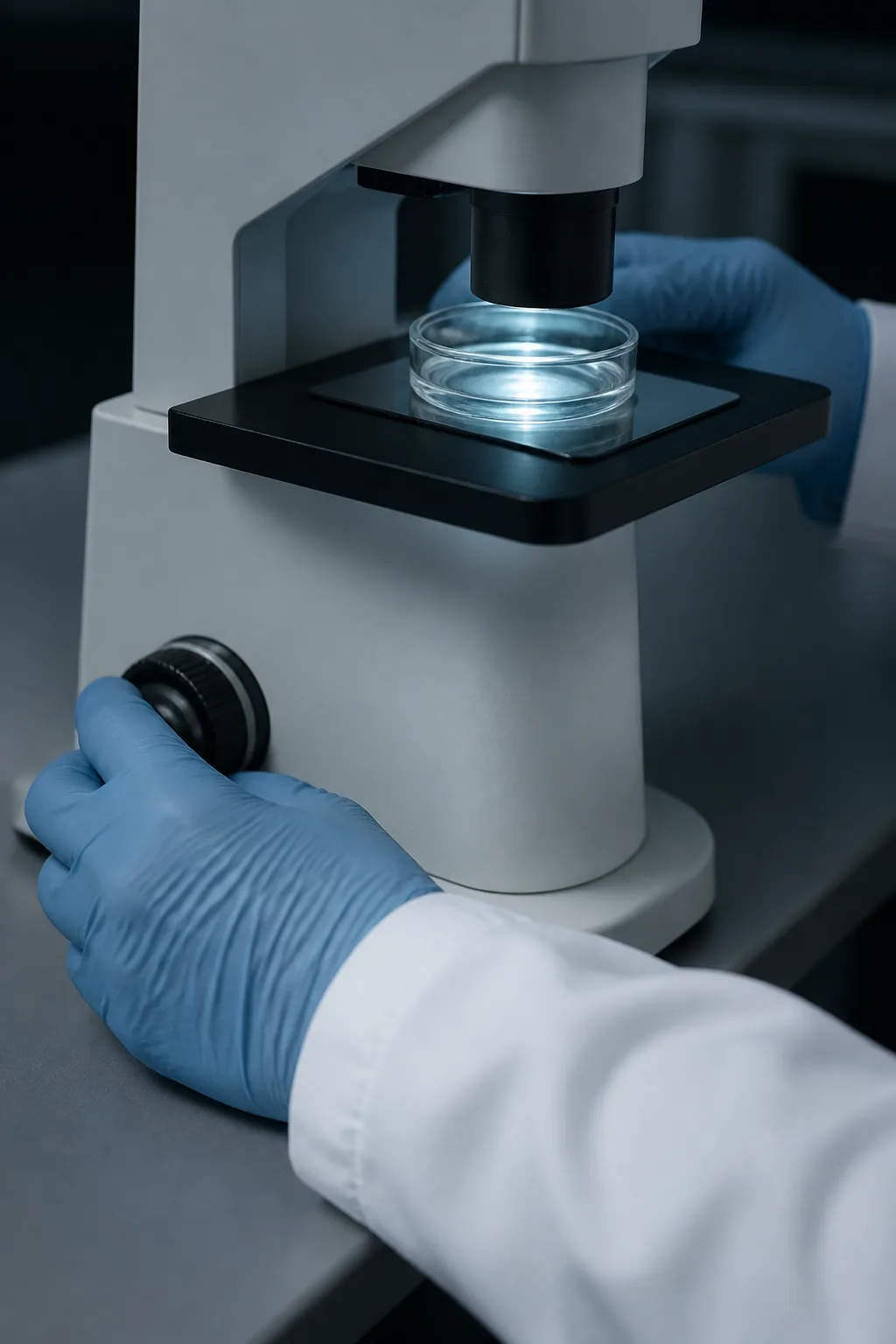
Plate B, about a forearm across: Watching. -
3.3 Staining
Dyes that mark one kind of cell, or one part of it, let us see which cells have stopped and where they sit.
What it can’t tell us No single stain says for certain that a cell has stopped; we look for several signs together.
-
3.4 Looking at shape
We trace outlines and compare sizes, because a stopped cell’s shape is one of its clearest signs.
What it can’t tell us A cell can be large for other reasons.
-
3.5 Modelling
We build sheets of cells in a computer, like Figure 1, to test whether an explanation hangs together before we try it in a dish. Sometimes a model you can hold makes a shape easier to think about.
What it can’t tell us A model contains only what we put in.
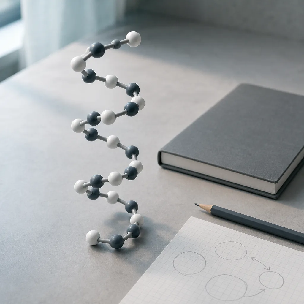
Plate C, about a hand across: A shape you can hold. The same shape as Figure 2 at two sizes: the helix in Figure 2 is a few millionths of a millimetre long; the model on the desk is about the length of a hand, tens of millions of times larger.


§4 · F5 of 10 · Midway
What have we found?
A paper usually puts its findings here.
§5 · F6 of 10 · Midway
What don’t we know yet?
We keep this list on the wall. It gets longer more often than it gets shorter.
-
Q1. What makes a resting cell decide not to wake?
A resting cell and a stopped one can look alike for a while; only following one cell for a long time tells them apart.
- Where we are
- Looking
- What would answer it
- Across, how long a cell has rested · up, how likely it is to start again (this is Figure 3)
-
Q2. Does a stopped cell change the cells around it, and how far does that reach?
A dish is crowded and busy; one cell’s influence is hard to separate from the rest.
- Where we are
- A guess we are testing
- What would answer it
- Across, distance from a stopped cell · up, how often its neighbours divide
-
Q3. Why does a stopped cell grow so large and flat?
Growing without dividing may be the cause of stopping, or only a side effect of it.
- Where we are
- Early
- What would answer it
- Across, time since it stopped · up, how much ground it covers
-
Q4. Is stopping one event, or many small ones that add up?
No single sign says for certain that a cell has stopped, so we look at several together.
- Where we are
- A guess we are testing
- What would answer it
- Across, small signs, one by one · up, whether the cell stops
-
Q5. Which of a cell’s proteins lose their shape first, and does it matter?
Shape is hard to see inside a living cell: an ordinary light microscope blurs anything smaller than about a fifth of a thousandth of a millimetre, and a protein is far smaller than that.
- Where we are
- Early
- What would answer it
- Across, how long ago a cell stopped · up, how many of its proteins keep their shape
-
Q6. Is “for good” always for good?
Stopping is thought to be permanent. We would like to know whether that is always true, and what it would take to see an exception.
- Where we are
- Looking
- What would answer it
- Across, time since stopping · up, chance of starting again
§6 · F7 of 10 · Late
What became of the cells you stained?
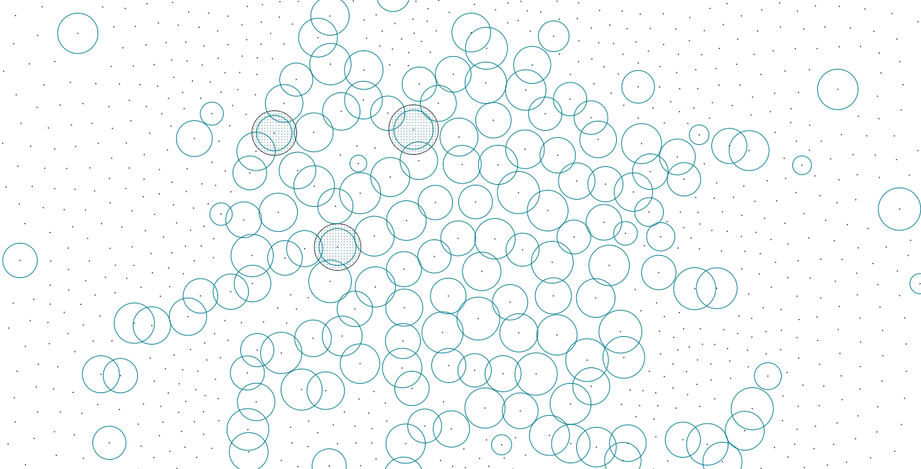
This is where the figure’s clock ends. In a dish, as in this drawing, stopped cells don’t leave. They stay, take up room and change what their neighbours do. Most of our questions start here.
§7 · F8 of 10 · Late
What is this lab not?
- We do not make products.
- We do not offer treatments or advice.
- We do not run trials, and we do not see patients.
- Nothing on this page is about making anyone live longer.
- The figures are illustrations of ideas, not results.
- If something sounds like a promise about your health, it did not come from us.
§8 · F9 of 10 · Late
Where is the work done?

Our rooms are in the city, on Kaurna Country. The day starts with whoever is on the morning check looking at every dish. Afternoons are long and quiet at the microscope. The model runs overnight, and in the morning we see what it drew. Students who want to know what this kind of research is like from the inside are welcome to write.

§9 · F10 of 10 · The end of the clock
How do you write to us?
Students looking for a project, researchers with a question in common, or readers who want to know more: write to us. We don’t give health advice.
A note on the type
The name at the top of this page is set in Martian Mono, a typeface that can widen and thin. Seven of its letters are tied to seven cells in Figure 1, and widen when their cell stops dividing. The headings are set on the same scale, one step later for each section, so the paper grows wider and thinner as you read it, the way the cells do. The scale is ours, drawn for this page. Prose is set in Geist. Figures 1, 2 and 4 were drawn in your browser as you read; Figure 3 is blank on purpose. There is no reference list: this page reports questions, not findings.
- Fig. 1 · A still
- Fig. 2 · A still
- Fig. 3 · Not drawn yet
- Fig. 4 · A still